|
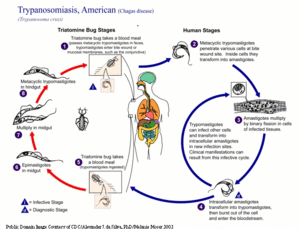
The life cycle of Trypanosoma cruzi involves both vertebrate and invertebrate hosts (see figure below). Metacyclic trypomastigotes are deposited on the mammalian (vertebrate host's) skin through the faeces of the triatomine bug vector. They have the capacity to penetrate skin through wounds, such as the bite from the bug, and across the mucosal membranes surrounding the eyes and mouth.
Inside the mammalian host, the trypomastigotes penetrate either phagocytic or non-phagocytic cells, in a manner distinct from phagocytosis. Parasites subvert the host cell Ca2+ -regulated lysosomal exocytic pathway, literally ‘hijacking’ lysosomes to enable them to invade effectively (Sibley and Andrews, 2000; Tan and Andrews, 2002). Within the host cell, trypomastigotes are initially held within a membrane bound vacuole. They subsequently enter the host cell cytoplasm directly, transforming into amastigotes (the intracellular replicative forms) (Tan and Andrews, 2002). Around five days post invasion, the amastigotes transform back into C- shaped trypomastigotes, and the host cell ruptures, releasing the parasites into the bloodstream. These bloodstream trypomastigotes can then either infect further cells, or can be taken up by a reduviid bug. Within the insect vector, epimastigotes develop in the alimentary tract, taking 10 – 15 days to replicate and transform into infective stages in the rectum (Kollien and Schaub, 2000). T. cruzi can also be transmitted via contaminated blood and infected organs used in transplant operations, or congenitally from mother to child.
|


